Kinetlas

The 3D anatomy atlas designed for learning the human body.

Explore human anatomy interactively. Study muscles, movements, systems, medical imaging and anatomical relationships from a new perspective.

1.664
anatomical structures
133
joint movements
43
animated exercises
4
languages

The whole human body.
On a single platform.

Every structure is a real 3D model you can rotate, isolate and study.

Muscles

289 muscles with origin, insertion, innervation and action.
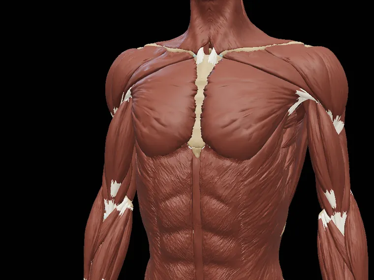
Trunk and arm muscles in the 3D model

Bones

159 bones and 911 bony landmarks on the model.
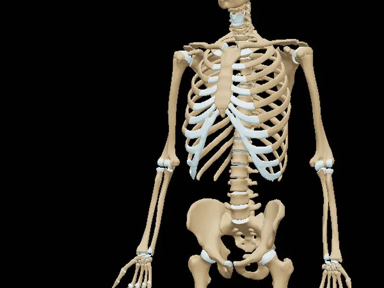
Skeleton in the 3D model

Joints

37 joints with ligaments, capsules and bursae.
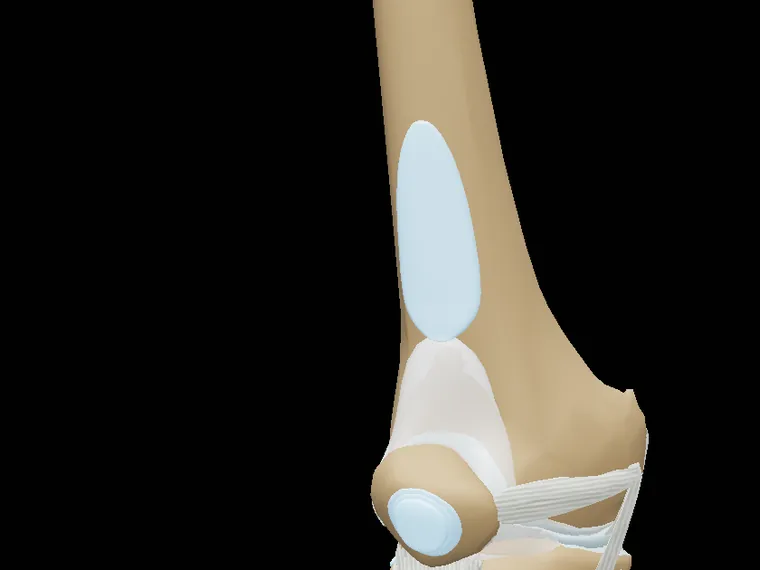
Knee ligaments in the 3D model

Systems

Nervous, cardiovascular, digestive, respiratory and more.
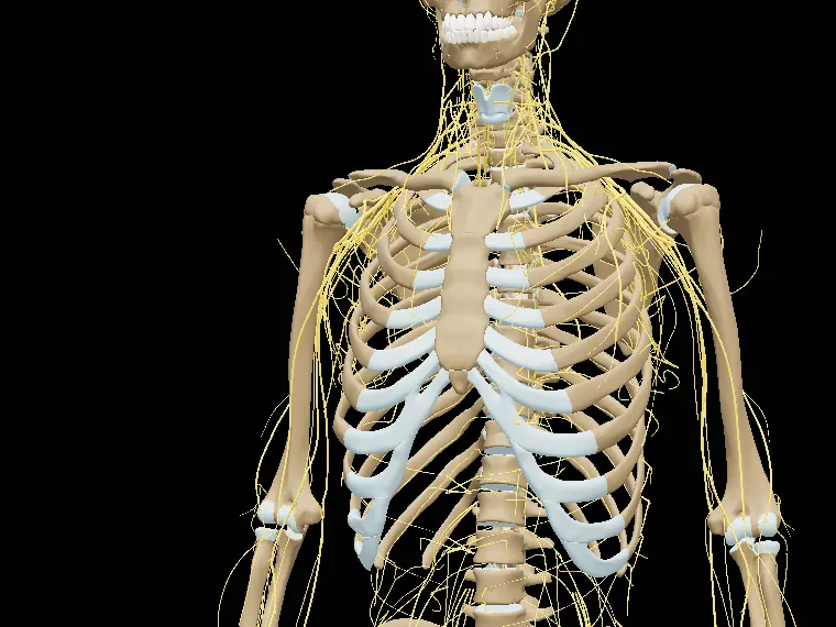
Peripheral nervous system over the skeleton

Biomechanics

133 movements with their axis, plane and muscles.
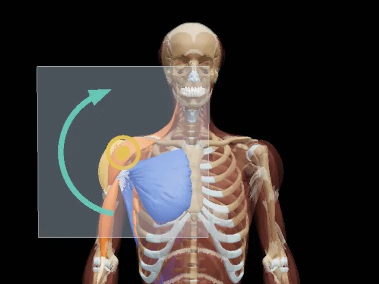
Shoulder abduction with its axis and plane of movement

Medical imaging

X-rays, CT and MRI from real studies.
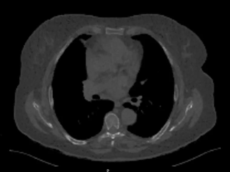
Sagittal slice of a head and neck CT

Dentistry

Every tooth in 3D, with its sections and dental anaesthesia.
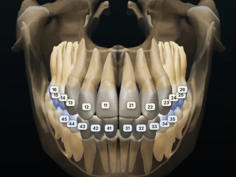
Numbered dental arches in the 3D model

Brain

Gyri, sulci and deep structures in their real colours.
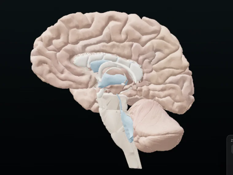
Sagittal section of the brain in the 3D model

Physiology

Muscle, bone and cartilage in 3D, level by level and step by step.
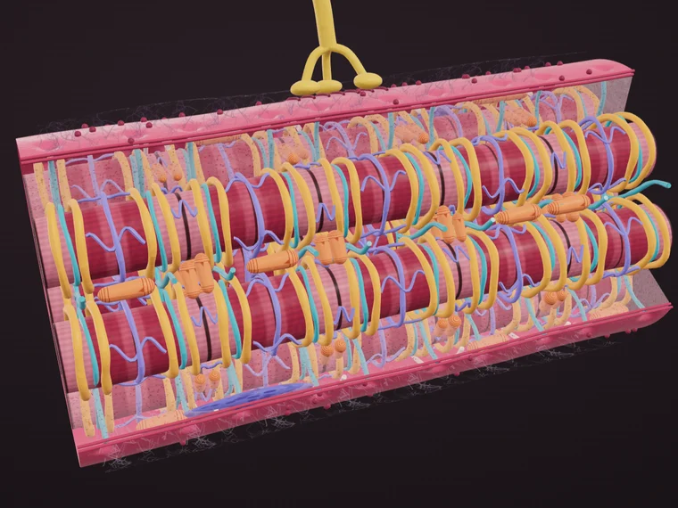
3D muscle fibre with its sarcolemma, T tubules and sarcoplasmic reticulum

Cross-sectional anatomy

3D slices of the body: go through each region layer by layer.
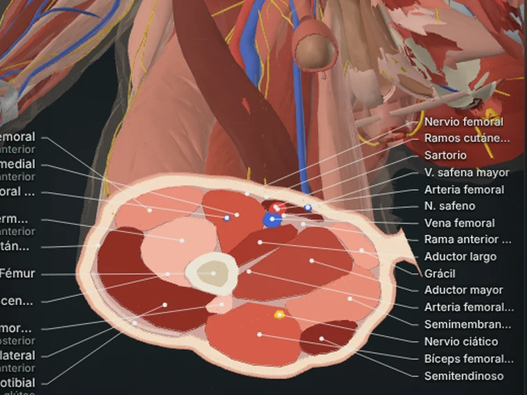
Cross-section of the thigh in the 3D model

Movement videos

Every muscle in action, with what it does in each movement.

Coming soon
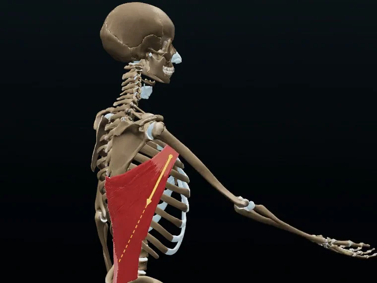
Video of the latissimus dorsi performing shoulder extension

Try it

Explore Kinetlas
without signing up.

Rotate the model and tap any bone or muscle. Your licence unlocks everything else.

Open the interactive 3D model

Open full screen

Muscles

Every muscle, explained in detail.

Select any muscle on the model and Kinetlas shows you what you need to know, from the essentials to the details.
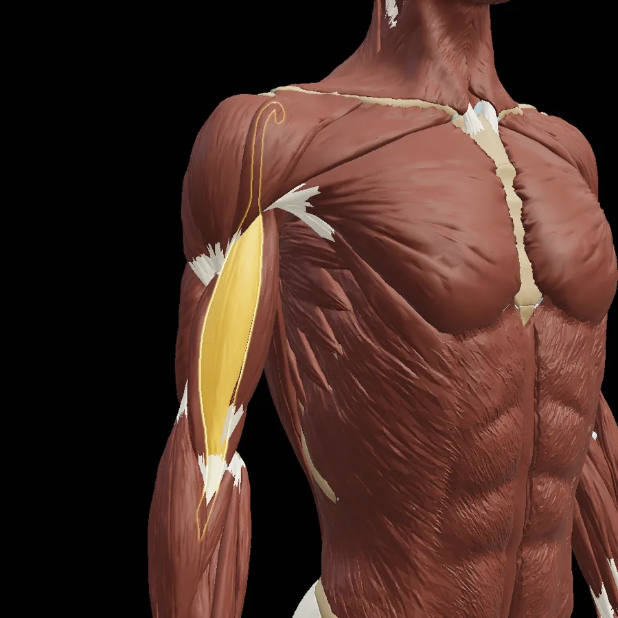
  • Origin and insertion, marked on the bone
  • Innervation with its roots, and main action
  • Movements it takes part in, and its role
  • Agonists, synergists and antagonists
  • Common injuries and exercises to strengthen it
Biceps brachii highlighted in the 3D model

Muscle · right

Biceps brachii, long head

Caput longum musculi bicipitis brachii

OriginSupraglenoid tubercle of the scapula
InsertionRadial tuberosity
InnervationMusculocutaneous n.C5–C6
ActionFlexion and supination of the forearm
Movements
Elbow flexion · Supination · Shoulder flexion
Agonists
Brachialis · Brachioradialis · Biceps, short head
Synergists
Pronator teres · Flexor carpi radialis
Antagonists
Triceps brachii · Anconeus
Relations
Bicipital groove · Musculocutaneous n. · Brachial a.
Injuries
Long head tendinopathy · SLAP lesion · Proximal rupture

Biomechanics

Not just what it is.
How it works.

Every movement with its axis and plane, and the muscles coloured by their role so you understand how they work together.
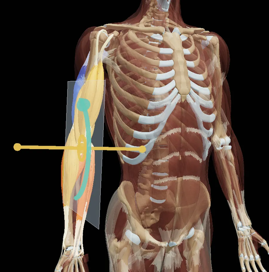
Elbow flexion with the muscles coloured by their role

Medical imaging

From anatomy to the clinical image.

X-rays, CT and MRI from real studies, organised by region, so you can recognise in the image what you study in 3D.
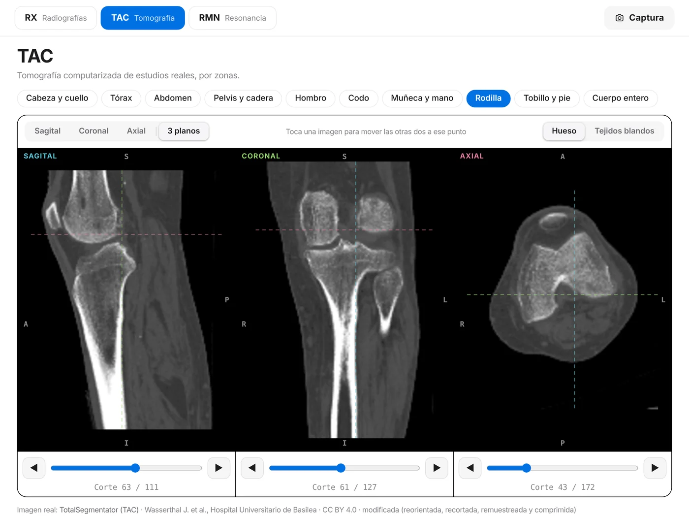
  • More than 1,300 real X-rays, of normal anatomy and pathology
  • X-rays with labelled structures to learn how to read them
  • CT of 10 regions and MRI of 6, each in its own section, slice by slice
  • Choose the plane: sagittal, coronal, axial or all three at once, synchronised
  • Bone window (default) and soft-tissue window on CT
  • A body figure always shows you where the slice is
Knee CT in Kinetlas with sagittal, coronal and axial slices at once
Real image: TotalSegmentator (CT), Wasserthal J. et al., University Hospital Basel · CC BY 4.0 · modified

Dentistry

The mouth, tooth by tooth.

A dedicated section to study the dentition in 3D, with the numbering you use and everything around it.
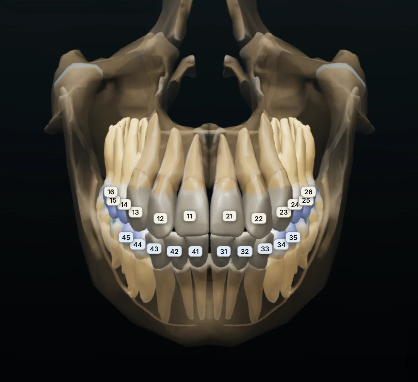
  • Permanent and deciduous dentition, with FDI numbering
  • Sections of each tooth to see enamel, dentine and pulp
  • Occlusal anatomy: cusps, fossae, grooves and ridges
  • 8 animated dental anaesthesia techniques, with the nerve they block
  • Nerves, gums and skull to place each tooth
Kinetlas dentistry section with the numbered dental arches

Physiology

From what it looks like
to how it works.

The physiology of the musculoskeletal system in 3D: go into the tissue level by level and see how it works step by step.

  • Muscle: from the whole muscle to the sarcomere, and contraction step by step
  • Bone: from the osteon to the osteocyte, and the remodelling cycle
  • Articular cartilage: its layers, how it bears load and what happens when it is damaged
  • Every structure with its name and its explanation
3D muscle fibre with its sarcolemma, T tubules and sarcoplasmic reticulum

Cross-sectional anatomy

Slice the body wherever you want.

Travel through the body from head to foot and see in each slice which muscles, vessels and nerves are there and how they relate.
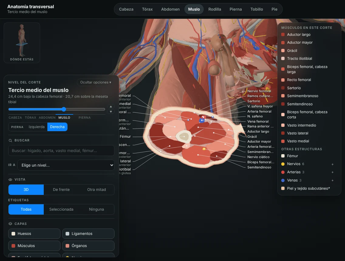
  • 3D slices of the head, thorax, abdomen, thigh, knee, leg, ankle and foot
  • Move the slice level and see how each structure changes
  • Labels, search and layers: bones, muscles, organs, nerves and vessels
  • A body figure shows you where the slice is, so you can relate it to CT and MRI
Kinetlas cross-sectional anatomy section with a slice of the thigh

Coming soon

Right now
we are filming.

We are filming videos of the movement of every muscle in the body and videos of injuries, so you can see how each muscle works and how each injury happens.

Videos of each muscle's movementInjury videos

They will be added to the platform as they are ready.

Features

Anatomy that makes sense.

Designed for Physiotherapy, Podiatry, Medicine and the other health sciences.

Study

Clear fact sheets for every structure, from the essentials to the details.

Review

Flashcards and multiple-choice questions generated from the atlas itself.

Understand relationships

What each nerve supplies, where each muscle inserts and what surrounds it.

Prepare for exams

Test yourself by region and by question type before the exam.

Connect anatomy and movement

Every muscle linked to the movements it produces or controls.

Learn visually

Rotate, isolate and make layers transparent until you understand each region.

For universities

A new way to teach anatomy.

An institutional version for health science faculties and schools, with access for all students and teaching staff.

  • Institutional licencesOne annual agreement for the whole faculty.
  • Student accessAccounts for each student, on any device.
  • Lecturer accessTo prepare classes and project in the classroom.
  • UpdatesContinuous improvements included in the licence.
  • New contentNew movements, exercises and regions every academic year.
  • Teaching toolsQuiz mode and class-ready views.
  • Lecturer feedbackContent is reviewed with teaching staff.
  • SupportDirect support throughout the academic year.

Licences

Choose how to use Kinetlas.

Personal licence

For students and professionals

Coming soon

Very soon you'll be able to activate it from the website.

  • Complete 3D atlas
  • Fact sheets, biomechanics and exercises
  • X-rays, CT and MRI
  • Dentistry
  • Quiz mode
  • Screenshots, notes, favourites and history

The licence is linked to your Kinetlas account.

Groups and universities

Licences for large groups

Contact us

Access for a whole class or faculty.

  • Access for students and lecturers
  • Updates and new content
  • Teaching tools
  • Support during the academic year
Contact us

We'll send you a proposal tailored to your institution.

We created Kinetlas as Physiotherapy students, with a simple idea: the atlas we wish we'd had to understand the body in motion.

Kinetlas · Barcelona

Start studying with Kinetlas.